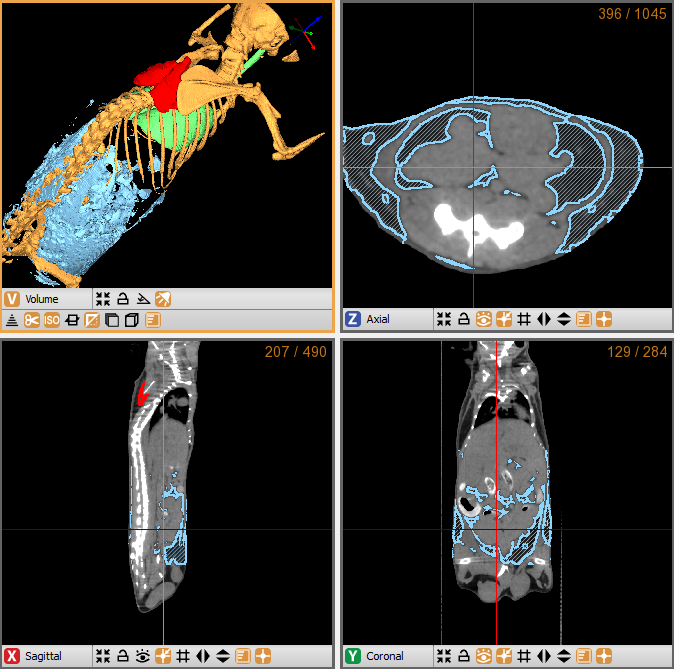
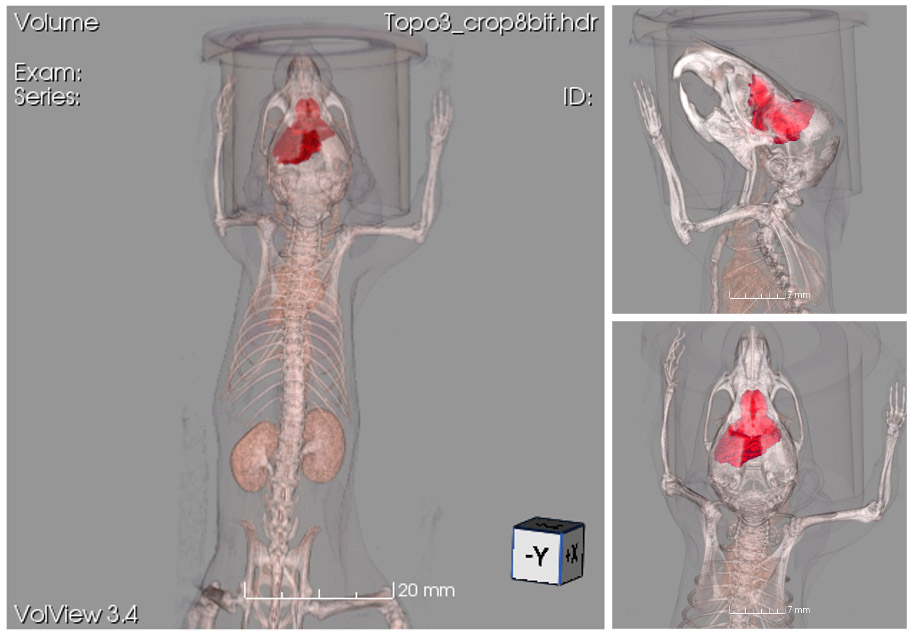
Research objective: Non-enhanced micro-CT image of a mouse, showing an example of quantitative segmentation (RED = Brown Adipose Tissue - BAT; CYAN = abdominal visceral white adipose tissue - WAT; GREEN - Lungs).
Animal model: Mouse, 20-25g
Acquisition protocol: 3 min acquisition time, 1 bed position, 55 kVp, 1 mA
Processing and reconstruction protocol: 0.08 mm isotropic voxel size
Biomarker or contrast agent: No tracer applied
Research objective: Rat CT imaging with contrast enhancement
Animal model: Wistar rat, ~300g
Acquisition protocol: 90 s acquisition time, 65 kVp, 1 mA, 1 bed position
Processing and reconstruction protocol: 0.06 mm isotropic voxel size
Biomarker or contrast agent: Continuous infusion of Iomeron(TM), 200 mgI/ml, 24 mL/h, 8 min infusion time; CT imaging time: 0 (basal), 4 min and 8 min after beginning of infusion (3D rendering refers to 4 min)
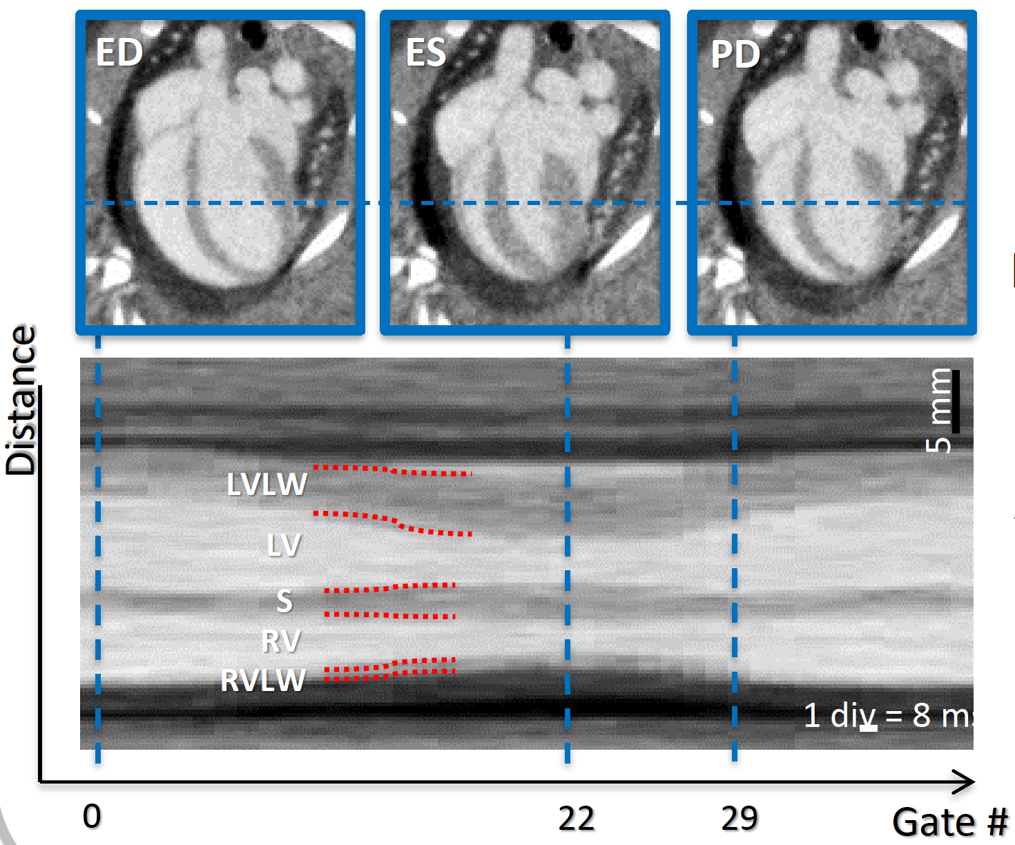
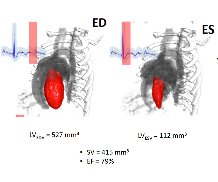
Research objective: Imaging and quantification of cardiac function. Extraction of M-Mode like MPR image to visualise and quantify Left Ventricle Lateral Wall (LVLW), Left Ventricle (LV), Septum (S), Right Ventricle (RV) and Right Ventricle Lateral Wall (RVLW). Segmentation of End-Diastolic (ED) and End-Systolic (ES) to quantify End Diastolic Volume (EDV) of left ventricle, End Systolic Volume (ESV) of left ventricle, Stroke Volume (SV) and Ejection Fraction (EF).
Animal model: Wistar rat, 480g
Acquisition protocol: 4 min acquisition time, 80 kVp, 1 mA, 20000 projections, 12 ms/proj
Processing and reconstruction protocol: 40 gates per R-R interval, 0.24 mm isotropic voxel size
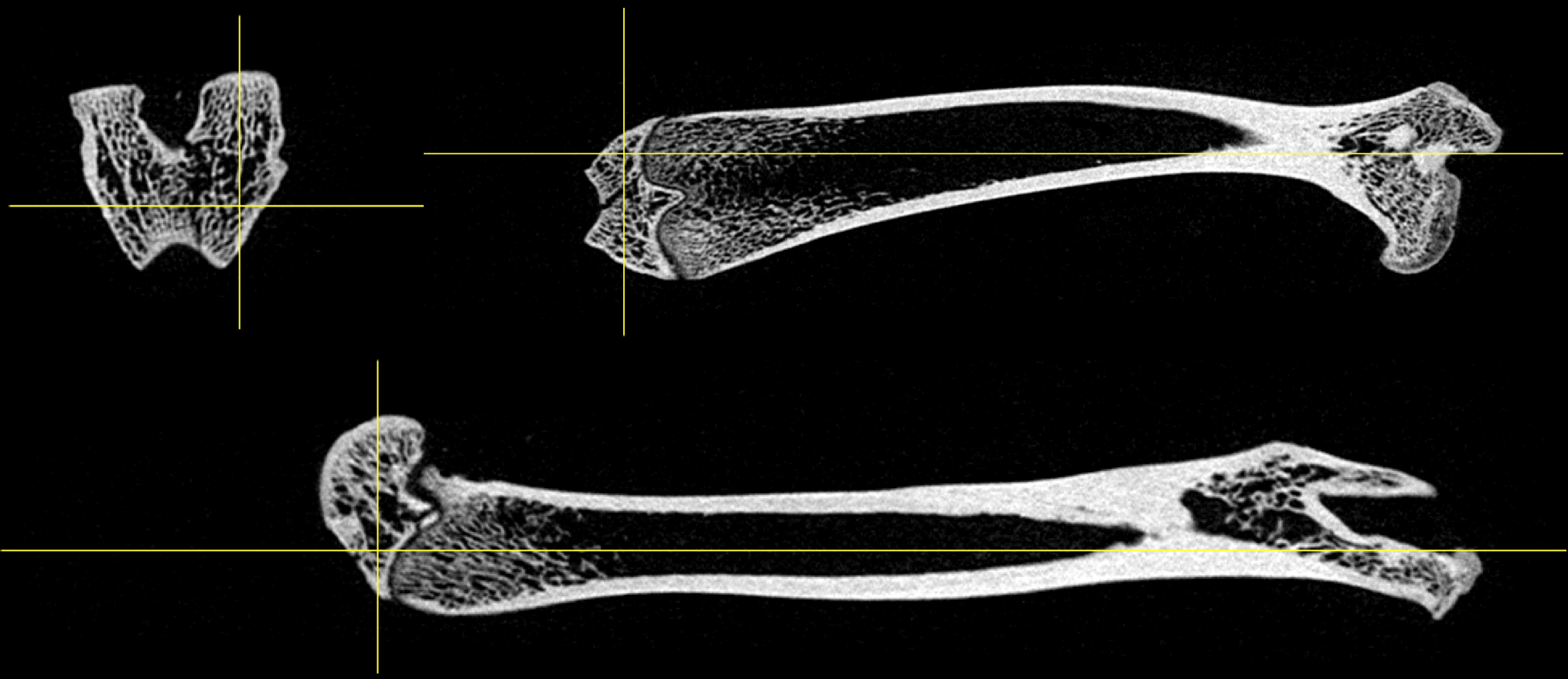
Research objective: Ex-vivo study for trabecula bone structure
Animal model: Rat femur bone
Acquisition protocol: 112 s acquisition time, 80 kVp, 1 mA, 2000 projections, 1 bed position
Processing and reconstruction protocol: 0.06 mm isotropic voxel size
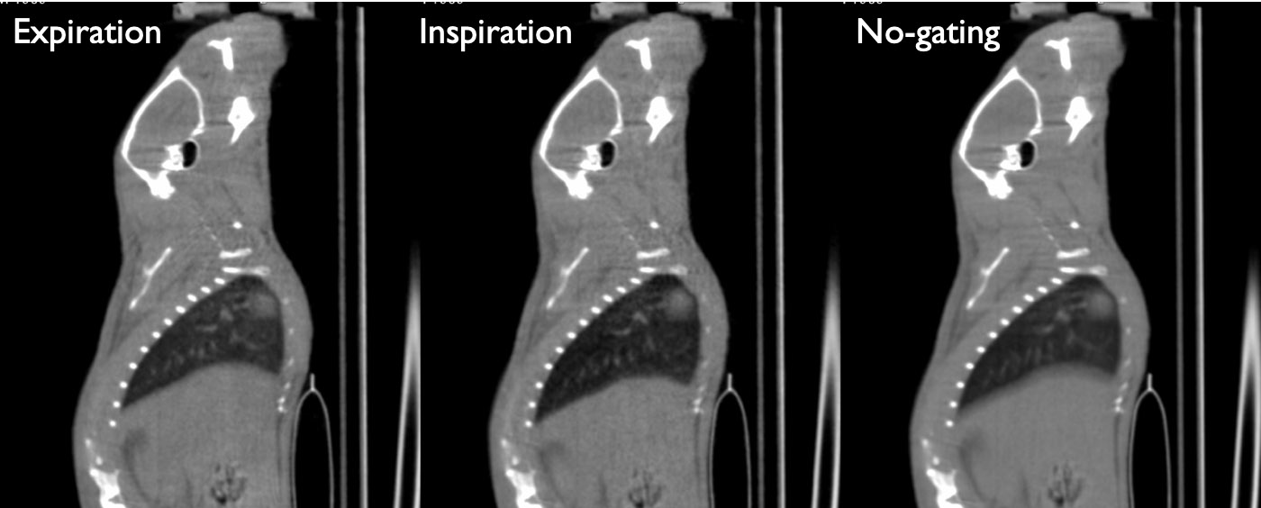
Research objective: Mouse CT lung imaging with automatic software-based gating, without using any hardware signal
Animal model: Mouse, ~25g, under anesthesia with isofluorane gas
Acquisition protocol: 90 s acquisition time, 65 kVp, 1 mA, 1 bed position
Processing and reconstruction protocol: Image processing into end-inspiration phase and end-expiration phase 0.16 mm isotropic voxel size
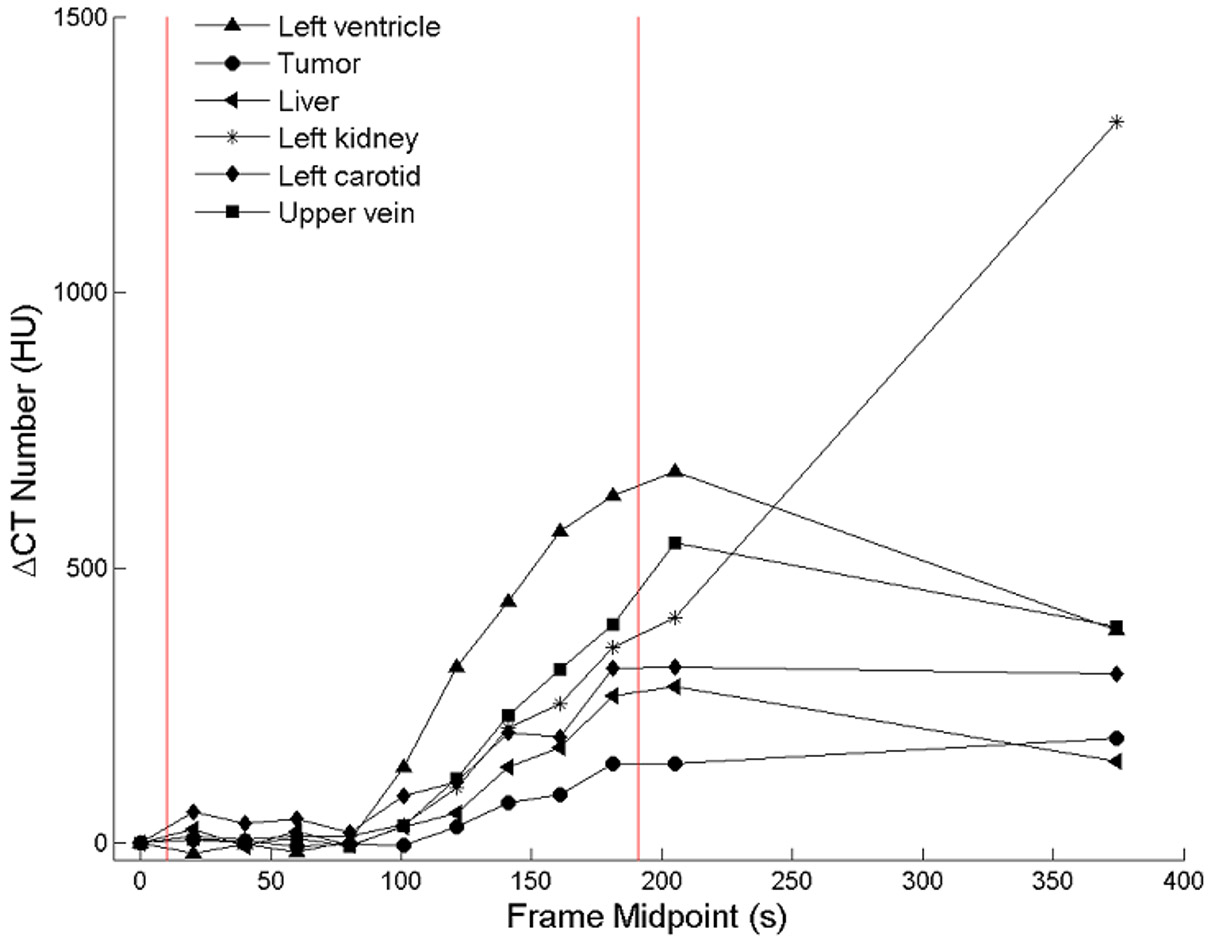
Research objective: Using 4D CT imaging to research morphology of mouse glioma and blood perfusion
Animal model: Mouse, ~25 g
Acquisition protocol: Scan 1 (4D): 7.3s acquisition time for each time frame, 80kV, 1mA, 10 frames in total. Scan 2 (3D): 40s total acquisition time, 80kV, 0.9mA.
Processing and reconstruction protocol: Scan 1: 0.24 mm isotropic voxel size. Scan 2: 0.12 mm isotropic voxel size.
Biomarker or contrast agent: Continuous infusion of Iomeron(TM), 200 mgI/ml, infusion speed 10 mL/h
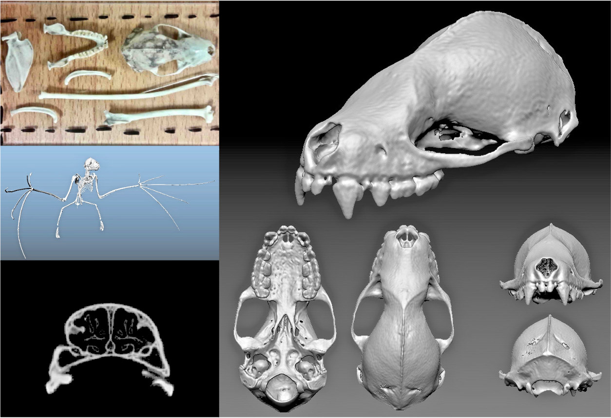
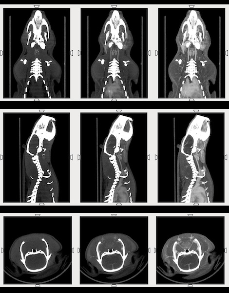
Research objective: Reconstitution of skeleton of Caribbean fruit bat
Animal model: Brachyphylla cavernarum. Dimensions: Skull length = 3 cm, Body length = 8.5 cm (incl. tail)
Acquisition protocol: 112 s acquisition time, 80 kVp, 1 mA, 1 bed position per plate, 6 acquisitions (complete skeleton), Total scan time < 12 min
Processing and reconstruction protocol: 0.06 mm isotropic voxel size